Did you know that diagnostic imaging IT, including patient scanning, diagnostics, tomography screening, and ultrasound scanning, is revolutionizing healthcare with its cutting-edge technology? From enhancing accuracy in diagnoses to improving patient outcomes, this innovative approach is reshaping the medical landscape. In today’s fast-paced world, the demand for efficient and precise diagnostic imaging solutions is at an all-time high. Leveraging the power of technology, diagnostic imaging IT streamlines processes, reduces errors, and ultimately saves lives. Stay tuned as we delve into the transformative impact of diagnostic imaging IT on healthcare delivery and patient care.
Key Takeaways
-
Understand the different imaging techniques used in diagnostic imaging to grasp their significance in healthcare.
-
Recognize the medical applications of diagnostic imaging,https://www.psmmis.com/diagnostic-imaging-it-specialists/ ranging from diagnostics to detecting diseases and monitoring treatment progress.
-
Explore specialized imaging methods that provide detailed insights into specific areas of the body, certain conditions, or diagnostics.
-
Prepare effectively for imaging appointments by following guidelines such as fasting or wearing appropriate clothing.
-
Stay informed about advancements in diagnostic imaging technology and diagnostics to make informed decisions about your healthcare.
-
Actively engage with healthcare providers to discuss the results and implications of diagnostic imaging tests.
Diagnostic Imaging Explained
Key Concepts
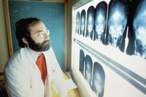
Diagnostic imaging is essential in healthcare for visualizing internal body structures to aid in disease diagnosis. It encompasses various medical imaging modalities like X-rays, CT scans, MRIs, and ultrasounds. These tests, conducted at diagnostic imaging centers, provide detailed images using imaging technique that help doctors identify and treat medical conditions effectively.
Different types of diagnostic imaging techniques cater to specific needs. X-rays are commonly used in medical imaging techniques studies to detect fractures, while CT scans offer detailed cross-sectional images useful for diagnosing tumors or internal injuries at diagnostic imaging centers. MRIs use magnetic fields to produce detailed images of soft tissues like the brain and muscles at diagnostic imaging centers.
Diagnostic imaging plays a crucial role in disease diagnosis and treatment planning. By providing clear visuals of organs and tissues, doctors at diagnostic imaging centers can pinpoint abnormalities early on. This aids in developing precise treatment strategies tailored to each patient’s condition, leading to better outcomes.
Importance in Healthcare
Diagnostic imaging is pivotal in modern healthcare due to its ability to detect diseases accurately and non-invasively. By utilizing advanced technologies, medical professionals can diagnose conditions at their early stages, increasing the chances of successful treatment outcomes.
The early detection facilitated by diagnostic imaging allows for timely interventions, potentially preventing disease progression and complications. For example, mammograms can detect breast cancer in its initial phases, enabling prompt treatment initiation and improving survival rates.
Moreover, diagnostic imaging significantly influences treatment outcomes and patient care. Through detailed imaging analysis, healthcare providers can monitor disease progression, evaluate treatment effectiveness, and make informed decisions regarding further interventions. This personalized approach enhances patient satisfaction and overall quality of care.
Imaging Techniques Overview
MRI Scans
MRI scans utilize magnetic fields and radio waves to generate detailed images of the body’s internal structures. These scans excel in visualizing soft tissues and organs, providing valuable insights for medical professionals. Common applications of MRI scans include diagnosing conditions such as tumors, injuries, and neurological disorders.
CT Scans
CT scans employ X-rays to produce cross-sectional images that offer a comprehensive view of the body’s anatomy. Known for their speed and accuracy, CT scans play a crucial role in detecting various medical conditions promptly. The benefits of CT scans extend to guiding treatment decisions and facilitating surgical planning processes.
Ultrasound
Ultrasound imaging relies on sound waves to create non-invasive images of internal body structures. Renowned for its versatility, ultrasound is capable of imaging different body parts, including the abdomen and heart. Moreover, ultrasound plays a pivotal role in monitoring fetal development during pregnancy.
X-rays
X-ray imaging operates on the fundamental principle of using electromagnetic radiation to produce images. With a rich historical significance, X-rays are widely recognized for their quick and painless imaging procedures. Commonly used in diagnosing fractures, infections, and various lung conditions, X-rays remain integral to medical diagnostics.
Mammography
Mammography serves as a critical tool in breast cancer screening, enabling early detection of abnormalities within breast tissue. By detecting breast irregularities at an early stage, mammograms significantly contribute to successful treatment outcomes. Guidelines recommend regular mammogram screenings based on age and individual risk factors.
Medical Uses of Imaging
Disease Diagnosis
Diagnostic imaging, including medical imaging and radiological imaging, is pivotal in detecting a myriad of diseases. Utilizing techniques like X-rays, CT scans, and MRIs, healthcare professionals can identify conditions such as fractures, tumors, and internal injuries accurately. The precision of these imaging methods aids in early detection and precise diagnosis.
Imaging plays a crucial role in swiftly and accurately diagnosing diseases, ensuring prompt initiation of treatment. Through medical photography and other imaging modalities, healthcare providers can visualize internal structures to pinpoint abnormalities effectively. This timely identification is essential for initiating appropriate medical interventions promptly.
Treatment Planning
In treatment planning, diagnostic imaging guides healthcare providers in formulating personalized treatment strategies. By analyzing images obtained from various medical imaging applications, physicians can evaluate disease progression and tailor treatment plans accordingly. These images provide valuable insights into the effectiveness of ongoing treatments.
Imaging tests are instrumental in monitoring treatment responses and adjusting therapeutic approaches as needed. By tracking changes in patients’ conditions through repeated imaging studies, healthcare professionals can gauge the efficacy of treatments. This dynamic approach ensures that interventions are modified based on real-time data, enhancing patient outcomes.
Deep Dive into Specific Techniques
MRI and MRA Scans
MRI and MRA scans serve distinct purposes in diagnostic imaging. MRI scans focus on detailed images of soft tissues, organs, and the brain, aiding in the detection of tumors, injuries, and neurological disorders. MRA scans, on the other hand, specifically target blood vessels to identify blockages or aneurysms that could lead to strokes or heart conditions.
The significance of MRI and MRA scans lies in their ability to provide non-invasive insights into vascular conditions and brain disorders. By capturing high-resolution images, these scans enable healthcare professionals to make accurate diagnoses and develop tailored treatment plans for patients suffering from a wide range of medical conditions.
CT Scan Insights
CT scans produce detailed cross-sectional images by combining multiple X-ray views. These scans offer rapid imaging capabilities, making them invaluable in emergency situations where quick diagnosis is crucial. In cases of trauma or internal bleeding, CT scans play a vital role in identifying injuries promptly and accurately.
The speed and precision of CT scans allow healthcare providers to swiftly assess traumatic injuries, internal bleeding, or other urgent medical conditions. This efficiency aids in expediting treatment decisions and improving patient outcomes by enabling timely interventions based on the scan results.
Ultrasound Applications
Ultrasound technology finds extensive applications across various medical specialties. From obstetrics to cardiology and musculoskeletal imaging, ultrasound plays a pivotal role in diagnosing conditions and guiding medical procedures. Its real-time imaging capabilities make it especially useful for visualizing internal structures during surgical interventions or biopsies.
The versatility of ultrasound extends to its ability to provide dynamic imaging for monitoring fetal development during pregnancy, evaluating heart function, or assessing soft tissue injuries. With its portability and non-invasive nature, ultrasound has become a cornerstone in modern medical diagnostics across different healthcare settings.
X-ray Significance
X-rays remain a fundamental tool in medical imaging due to their ability to penetrate tissues and create detailed images of bones and organs. In diagnosing skeletal injuries or pulmonary conditions like pneumonia, X-rays offer quick insights into the extent of damage or abnormalities present.
Advancements in X-ray technology have enhanced imaging quality while minimizing radiation exposure risks for patients. With digital X-ray systems and advanced image processing techniques, healthcare providers can now obtain clearer images with reduced radiation doses, ensuring safer diagnostic procedures.
Specialized Imaging Methods
Bone Density Scans
Bone density scans are crucial in evaluating bone health, detecting osteoporosis, and monitoring treatment effectiveness. These tests utilize low-dose X-rays to measure bone mineral density. By assessing bone strength, they help prevent fractures and assess fracture risk accurately.
Early detection of bone density loss through screening is vital for timely intervention and management. It enables healthcare providers to implement preventive measures and lifestyle changes effectively. Regular screenings empower individuals to take proactive steps towards maintaining optimal bone health.
Arthrogram Procedures
Arthrograms are specialized imaging techniques used for evaluating joint conditions. They involve injecting a contrast agent into the joint space before imaging. This process enhances the visualization of soft tissues, ligaments, and cartilage within the joint.
Combining imaging with contrast agents in arthrograms provides detailed insights into joint injuries, tears, and inflammatory conditions. This technique aids physicians in accurate diagnosis and formulation of appropriate treatment plans for patients with joint-related issues.
Myelogram Tests
Myelogram tests play a crucial role in assessing spinal cord and nerve root abnormalities. During this procedure, a contrast dye is injected into the spinal canal to highlight spinal structures on X-ray images. It helps in identifying spinal tumors, herniated discs, or other spinal pathologies accurately.
The diagnostic value of myelogram tests lies in their ability to detect subtle spinal abnormalities that may not be visible on standard imaging studies. By providing detailed images of the spinal cord and nerve roots, these tests aid healthcare professionals in making precise diagnoses and planning appropriate interventions.
Preparing for Imaging Appointments
Scheduling Tips
Patients should schedule diagnostic imaging appointments well in advance to secure preferred dates and times. It’s crucial to confirm the appointment details, including the type of imaging test and any specific preparation instructions. Following the facility’s guidelines ensures accurate results.
Before specific imaging tests like MRI or CT scans, patients may need to fast for a certain period. Avoiding food or drinks before the appointment helps obtain clear images. Informing the healthcare provider about any metal implants or devices is essential for safety during MRI scans.
Adhering to scheduling instructions is vital for optimal imaging results. Patients should arrive on time for their appointments and bring all necessary documents, such as insurance cards and identification. Rescheduling may be required if preparation steps are not followed accurately.
What to Expect
During X-rays, patients can expect to feel slight discomfort due to positioning requirements. However, the procedure is quick and painless. For ultrasounds, a gel is applied to the skin, which might feel cold but causes no discomfort during the scan.
In MRI scans, patients may experience loud noises from the machine but are provided with earplugs for protection. The technician will guide patients throughout the process, ensuring comfort and safety. CT scans involve lying still on a table that moves through a ring-shaped scanner, capturing detailed images.
Patients undergoing imaging tests should not worry about radiation exposure as modern machines emit minimal levels deemed safe by healthcare standards. Any concerns regarding discomfort or claustrophobia can be addressed with the healthcare provider beforehand.
Staying Informed
Latest News
Diagnostic imaging technology has recently witnessed remarkable advancements, enhancing the accuracy and efficiency of medical diagnoses. Cutting-edge techniques like 3D mammography and artificial intelligence integration are revolutionizing the field. These innovations enable healthcare professionals to detect abnormalities with greater precision, leading to early intervention and improved patient outcomes.
In a groundbreaking study published in 2021, researchers demonstrated how AI algorithms can analyze medical images to identify subtle patterns indicative of various diseases. This development not only expedites the diagnostic process but also reduces the chances of human error, ensuring more reliable results. As a result, patients receive timely and accurate diagnoses, paving the way for more effective treatment plans.
The introduction of portable ultrasound devices has significantly transformed diagnostic imaging practices, especially in remote or underserved areas. These handheld devices provide healthcare providers with on-the-spot imaging capabilities, enabling quick assessments and immediate interventions. This portability enhances accessibility to diagnostic services, particularly benefiting patients in rural communities or emergency situations.
Recent Conferences
The recent Diagnostic Imaging Conference held in 2022 brought together experts from around the world to discuss the latest trends and challenges in the field. Key discussions centered on the integration of machine learning algorithms into imaging technologies, streamlining workflows and improving diagnostic accuracy. The exchange of knowledge and experiences at such conferences is crucial for driving innovation and advancing patient care.
Emerging trends in diagnostic imaging include the utilization of virtual reality (VR) for surgical planning and training purposes. By creating immersive 3D models from medical images, VR technology allows surgeons to visualize complex procedures beforehand, enhancing surgical precision and reducing risks. This innovative approach not only benefits healthcare professionals but also contributes to better surgical outcomes for patients.
Collaboration among radiologists, technologists, and other healthcare professionals is essential for staying abreast of developments in diagnostic imaging. By sharing insights and best practices, professionals can enhance their skills and adapt to evolving technologies effectively. Knowledge sharing fosters a culture of continuous learning and improvement, ultimately benefiting patients by ensuring they receive the highest quality care possible.
Final Remarks
You’ve gained valuable insights into the world of diagnostic imaging, understanding its various techniques, medical applications, and preparation procedures. By staying informed and proactive about your imaging appointments, you take control of your health journey. Remember, knowledge is power when it comes to making informed decisions about your well-being.
Ensure you ask questions during your appointments, follow the preparation guidelines diligently, and stay proactive in seeking out the best imaging options for your needs. Your commitment to understanding and engaging with diagnostic imaging not only benefits your health but also empowers you to be an active participant in your healthcare decisions. Stay informed, stay proactive, and prioritize your health above all else.
Frequently Asked Questions
What is diagnostic imaging and why is it important?
Diagnostic imaging involves using various technologies to produce images of the body’s internal structures, aiding in the diagnosis and treatment of medical conditions. It is crucial for detecting diseases early, guiding interventions, and monitoring treatment progress.
How do different imaging techniques vary in their applications?
Imaging techniques vary in how they capture images of the body, such as X-rays, CT scans, MRIs, ultrasounds, and PET scans. Each technique has specific strengths in visualizing different types of tissues or functions within the body, allowing for comprehensive diagnostic evaluations.
What are the common medical uses of diagnostic imaging?
Diagnostic imaging is commonly used to assess injuries, detect tumors or abnormalities, monitor the effectiveness of treatments, guide surgical procedures, and screen for diseases like cancer or heart conditions. It provides valuable insights for healthcare providers to make informed decisions about patient care.
How should one prepare for an imaging appointment?
Before an imaging appointment, individuals may need to fast, avoid certain medications, wear comfortable clothing without metal objects, and inform the technologist about any relevant medical history or conditions. Following these preparation guidelines helps ensure accurate and efficient imaging results.
What are some specialized imaging methods available?
Specialized imaging methods include functional MRI (fMRI) for brain activity mapping, nuclear medicine scans for metabolic processes, angiography for blood vessel visualization, and elastography for tissue stiffness assessments. These advanced techniques offer detailed insights into specific aspects of anatomy and physiology for precise diagnoses.